Call Us Today!
(952) 465-0105
New Patients
(952) 225-5280


Wisdom teeth, clinically known as third molars, are the last teeth to develop and usually appear between the late teens and mid-twenties. Because they erupt later than other teeth, they often encounter limited space in the jaw and unusual angulation as they try to come in. Not every wisdom tooth causes trouble, but their unpredictable timing and position mean they deserve careful evaluation rather than a casual “wait and see” approach.
When a wisdom tooth cannot erupt fully or aligns improperly, it can create pressure, inflammation, or pockets that trap food and bacteria. This environment increases the likelihood of localized infection, gum irritation, and decay on the wisdom tooth or its neighbors. Because these effects can progress quietly, regular clinical and radiographic monitoring is essential to identify problems early and preserve long-term oral health.
Our team treats wisdom teeth with an emphasis on individualized care. We combine a detailed clinical exam with targeted imaging to determine whether a tooth is likely to erupt normally or if intervention is the safer course. By assessing each patient’s anatomy, dental development, and overall health, we help families and adults make informed decisions about observation versus removal.
Many people first notice an issue with their wisdom teeth through pain or swelling at the back of the mouth. Pain can be intermittent or persistent and may worsen with chewing. Swelling and redness of the gum tissue around a partially erupted tooth are common signs of infection; this presentation often coincides with bad taste or difficulty opening the mouth fully.
Other indicators include recurrent decay on the wisdom tooth or on the adjacent second molar, unexplained shifting of nearby teeth, and the formation of fluid-filled sacs called cysts. In some cases, impacted third molars can contribute to gum disease in the surrounding area or damage the roots of neighboring teeth. Because symptoms can vary widely, even subtle changes warrant professional assessment.
It’s also possible for problematic wisdom teeth to be asymptomatic early on, which is why preventive evaluation matters. Routine dental exams and targeted x-rays allow clinicians to spot developing impactions or risky positions before pain or damage occurs. When early warning signs are found, planning a controlled, timely intervention can simplify the procedure and recovery.
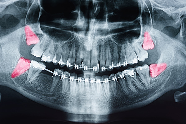
A careful diagnosis starts with a thorough medical and dental history followed by a focused intraoral exam. We routinely use panoramic radiographs to view all third molars and their relationship to the jaw, and we employ 3D imaging when finer detail is needed. Three-dimensional x-rays provide precise information about root configuration, bone density, and proximity to critical structures such as the inferior alveolar nerve or the maxillary sinus.
Classification of an impaction—soft tissue versus bony, vertical versus angled—helps guide the treatment strategy. Soft tissue impactions involve a tooth covered primarily by gum tissue, while bony impactions are partially or fully encased in jawbone. Each pattern has practical implications for surgical access, anticipated healing, and the type of anesthesia that will best ensure patient comfort.
After gathering diagnostic data, our surgeons discuss the findings and recommended options in plain language. We explain what the imaging reveals, outline the benefits and risks of removing versus monitoring a tooth, and describe the expected recovery process. This shared decision-making approach helps patients and families choose the plan that fits their health profile and personal priorities.
Wisdom tooth removal ranges from straightforward extractions to more involved surgical procedures, depending on the tooth’s position and root anatomy. Procedures are performed in a calm, controlled environment with appropriate monitoring, and anesthesia is tailored to each patient’s needs. Options include local anesthesia, nitrous oxide, intravenous (IV) sedation, or general anesthesia—choices we discuss in advance so you know what to expect.
When a tooth is impacted, the surgeon may need to gently remove some bone and section the tooth into smaller pieces for safe removal. These steps are routine and are done with techniques that prioritize precision and tissue preservation. Throughout the procedure, our goal is to minimize trauma and to protect adjacent teeth, nerves, and sinus structures while achieving complete, hygienic removal of the problematic tooth.
Before you leave the office we review post-procedure instructions, arrange any necessary prescriptions, and schedule follow-up care. Our surgeons place a premium on patient comfort and safety, and we make sure you have clear guidance on wound care, dietary recommendations, and signs that warrant a prompt return to the office.
Initial healing after extraction typically spans several days, with most patients experiencing peak swelling and discomfort in the first 48–72 hours. Managing swelling with cold packs, following a soft-food diet, and taking prescribed or recommended pain medications help patients stay comfortable while soft tissues begin to close. Rest and limited physical exertion during the first few days support optimal healing.
Certain complications are possible—such as dry socket, infection, or temporary nerve sensitivity—but they are well-understood and manageable when identified early. We provide clear instructions on oral hygiene, activity limitations, and warning signs to watch for, and we make follow-up visits available to monitor healing. If antibiotics or additional interventions are needed, those decisions are guided by clinical findings and best-practice protocols.
For younger patients, early treatment can simplify recovery because root development and bone flexibility are generally more favorable. For older patients or those with complex medical histories, the surgical plan is adapted to reduce risk and support predictable outcomes. In all cases, careful planning, skilled technique, and attentive follow-up help minimize complications and restore oral health efficiently.
At Northern Star Oral and Maxillofacial Surgery LLC, our team approaches wisdom tooth care with a balance of technical expertise and patient-centered communication. If you or a family member is experiencing symptoms, or if routine imaging has raised questions about third molars, contact us to discuss your situation and learn more about the evaluation and treatment options available. We are happy to explain what to expect and help you choose the path that best protects your long-term oral health.
Wisdom teeth, also called third molars, are the last teeth to develop and typically appear in the late teens to mid‑twenties. Because they erupt after most other teeth, they often encounter limited space or atypical angulation as they emerge. Not every wisdom tooth causes problems, but their late timing and variable positions make careful evaluation important.
When a wisdom tooth cannot erupt fully or presses against neighboring teeth it can create pockets that trap food and bacteria, increasing the risk of infection and decay. Impacted teeth can also contribute to gum inflammation, cyst formation, or shifting of adjacent teeth. Regular clinical and radiographic monitoring helps identify early signs of trouble so treatment can be planned proactively.
Common symptoms include pain or tenderness at the back of the mouth, swelling and redness of surrounding gum tissue, and a persistent bad taste or bad breath. Some patients experience difficulty opening the mouth fully or pain that worsens with chewing, which can indicate infection or inflammation. Symptoms may be intermittent at first and can escalate if underlying issues are not addressed.
Other indicators include recurrent decay on a wisdom tooth or the adjacent second molar, unexplained shifting of nearby teeth, and the development of fluid‑filled sacs called cysts. In many cases problematic wisdom teeth are asymptomatic early on, so radiographic evaluation is important even when discomfort is minimal. Timely assessment allows for simpler, more predictable treatment when intervention is needed.
Evaluation begins with a thorough medical and dental history and a focused intraoral examination to assess eruption, gum health, and signs of infection or decay. Panoramic radiographs are routinely used to view all third molars and their relationship to surrounding structures, and three‑dimensional imaging may be employed when more detail is required. Imaging reveals root shape, bone density, and proximity to critical anatomy such as the inferior alveolar nerve or the maxillary sinus.
Classification of an impaction—soft tissue versus bony, vertical versus angled—helps guide the recommended approach and anesthesia needs. The surgeon considers the patient’s age, overall health, dental development, and symptoms when weighing observation against removal. We use plain, evidence‑based explanations so patients and families can make informed, shared decisions about the plan of care.
Extraction is typically recommended for wisdom teeth that are symptomatic, repeatedly infected, causing decay to themselves or adjacent teeth, contributing to gum disease, or associated with cysts or tumors. Removal may also be advised when a tooth’s angulation or impaction is likely to damage neighboring roots or compromise future dental work. In orthodontic cases, third molar removal can be part of a broader treatment plan to preserve alignment.
Observation is a reasonable option when a wisdom tooth is fully erupted, functional, free of pathology, and easily cleaned. Patients under observation should have regular clinical exams and targeted x‑rays so clinicians can detect changes early. Because younger patients often have more favorable root development and bone flexibility, timely removal when appropriate can simplify the procedure and recovery.
Anesthesia choices include local anesthesia, nitrous oxide (laughing gas), intravenous (IV) sedation, and general anesthesia, and the selection depends on the complexity of the extraction and the patient’s comfort and medical history. Local anesthesia numbs the surgical site while sedation or general anesthesia provide varying degrees of relaxation or unconsciousness for greater comfort during more invasive procedures. Our team discusses anesthesia options in advance and matches the plan to each patient’s needs and safety considerations.
All anesthesia types are administered with appropriate monitoring by trained staff and follow established safety protocols to ensure a controlled environment. Preoperative instructions—such as fasting guidelines for certain sedation levels and medication adjustments—are provided to reduce risk and optimize outcomes. Clear communication about these instructions helps patients prepare and reduces the chance of complications on the day of surgery.
Surgical extraction typically begins with administration of the chosen anesthesia and careful monitoring. The surgeon makes a small incision in the gum to expose the tooth, removes any necessary bone, and may section the tooth into smaller pieces to facilitate gentle removal. These steps are routine and performed with instruments and techniques designed to preserve surrounding tissues and minimize trauma.
After the tooth is removed the socket is irrigated, the area is inspected for debris or sharp edges, and sutures may be placed to support healing and reduce bleeding. The surgeon reviews postoperative care instructions and arranges any necessary prescriptions and follow‑up visits before you leave. Emphasis is placed on protecting nearby nerves and sinus anatomy throughout the procedure to reduce the risk of complications.
Initial healing usually involves peak swelling and discomfort during the first 48–72 hours, which can be managed with cold packs, prescribed or recommended analgesics, and a soft‑food diet. Rest and avoidance of vigorous physical activity during the early days support optimal healing, while gentle oral hygiene helps prevent infection. Smoking and use of straws should be avoided because suction can disturb the surgical site and increase the risk of dry socket.
Follow the specific wound care and oral hygiene instructions provided by your surgeon, including when to resume brushing and how to irrigate the area if advised. Attend scheduled follow‑up appointments so the team can monitor healing and address any concerns promptly. Contact the office promptly for signs such as uncontrolled bleeding, fever, severe or increasing pain, or altered sensation that persists beyond the expected recovery window.
Known complications include dry socket (alveolar osteitis), postoperative infection, prolonged bleeding, and temporary nerve disturbance causing numbness or tingling. Dry socket typically presents as intense pain a few days after extraction and is treated with local measures, medicated dressings, and pain control; infections are managed with drainage and antibiotics when indicated. Temporary nerve changes usually resolve over weeks to months, but the risk is assessed preoperatively based on imaging and surgical complexity.
Our approach emphasizes prevention through careful planning, gentle surgical technique, and attentive postoperative guidance. When complications occur they are managed promptly using evidence‑based protocols, and patients are followed closely until the issue resolves. Early communication with the surgical team improves the ability to treat complications effectively and minimize long‑term effects.
When medical conditions or medications are present, the surgeon coordinates care with the patient’s medical providers as needed and adjusts perioperative instructions to ensure safety. Preoperative screening, appropriate imaging, and a conservative surgical approach all contribute to predictable outcomes across age groups. Individualized planning ensures that each patient receives the safest, most appropriate care for their situation.
To schedule an evaluation, contact Northern Star Oral and Maxillofacial Surgery LLC in St. Louis Park to discuss symptoms or concerns and arrange a convenient appointment time. New patients may call the dedicated new‑patient line and established patients can use the existing‑patient number listed on the practice homepage for phone scheduling and previsit instructions.
At the evaluation the surgeon will review your medical and dental history, perform a focused exam, and obtain any necessary radiographs to develop a personalized recommendation for observation or treatment. The practice emphasizes clear communication and shared decision making so you understand the findings, options, and expected recovery before proceeding.